Fundus Fluorescein Angiography
OverView
FFA stands for Fundus Fluorescein Angiography, a diagnostic procedure used in ophthalmology to examine the circulation of the retina and choroid (the layers at the back of the eye). This test is particularly useful for detecting and monitoring various eye conditions, especially those related to the retina, such as diabetic retinopathy, age-related macular degeneration (AMD), and retinal vein occlusions.
How Fundus Fluorescein Angiography Works
Fluorescein Dye Injection:
- A yellow fluorescein dye is injected into a vein in the patient's arm. The dye travels through the bloodstream and reaches the blood vessels in the retina and choroid.
Image Capture:
- As the dye circulates through the blood vessels in the eye, a series of photographs is taken using a special camera equipped with filters. These filters allow the camera to capture the fluorescence emitted by the dye as it illuminates the blood vessels.
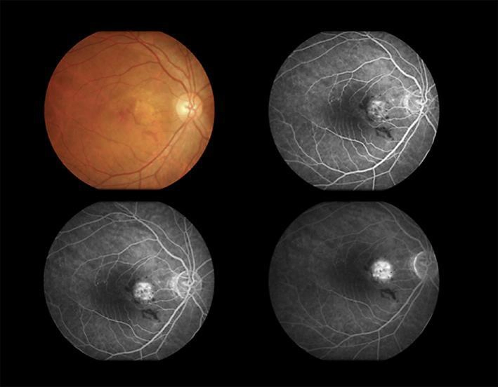
Phases of Angiography:
- The procedure typically captures images in different phases —early, mid, and late phases—to observe how the dye fills the blood vessels and how it leaks or pools in the retinal tissues.
Analysis of Results:
- The resulting images provide detailed information about the blood flow in the retina and choroid, allowing the ophthalmologist to identify abnormalities such as blockages, leaks, or areas of non-perfusion (where blood flow is absent).
Indications for FFA
Fundus fluorescein angiography is indicated for diagnosing and monitoring several retinal and choroidal conditions, including:
Diabetic Retinopathy:
- FFA can detect microaneurysms, neovascularization (new, abnormal blood vessels), and areas of retinal ischemia (lack of blood supply) in patients with diabetes.
Age-Related Macular Degeneration (AMD):
- It helps in identifying wet AMD, where abnormal blood vessels leak fluid or blood into the macula, leading to vision loss.
Retinal Vein Occlusion:
- FFA is used to assess the extent of blockage in the retinal veins and to detect areas of retinal ischemia and edema (swelling).
Retinal Detachment:
- The procedure can help identify the location and extent of retinal detachment, particularly in cases where the detachment is caused by a tear in the retina.
Choroidal Neovascularization:
- FFA helps detect abnormal blood vessels growing under the retina, often associated with conditions like AMD and myopic degeneration.
Macular Edema:
- The test can pinpoint areas of fluid accumulation in the macula, which can occur in various retinal diseases.
The FFA Procedure
Preparation:
Pupil Dilation:
- The patient's pupils are dilated with eye drops to allow a better view of the retina.
Injection:
- A small needle is used to inject fluorescein dye into a vein, usually in the arm or hand. Some patients may feel a brief warm sensation as the dye enters the bloodstream.
Image Capture:
- The patient is seated with their chin on a chin rest, and the camera is aligned with the eye. A series of photographs is taken over a period of several minutes as the dye passes through the retinal and choroidal blood vessels.
Post-Procedure:
- The patient’s skin and urine may appear slightly yellow for a short time after the procedure as the dye is excreted from the body. This is normal and usually resolves within 24-48 hours.
- Patients are typically advised to avoid bright light for a few hours due to the dilation of the pupils.
Benefits of FFA
Detailed Visualization:
- FFA provides detailed images of the retinal and choroidal vasculature, allowing for precise identification of abnormalities.
Early Detection:
- The procedure can detect changes in the blood vessels of the retina and choroid before they are visible on a standard retinal examination, enabling early intervention.
Guiding Treatment:
- The results of FFA can help guide treatment decisions, such as the need for laser therapy, intravitreal injections, or surgery.
Risks and Considerations
Allergic Reactions:
- While rare, some patients may experience an allergic reaction to the fluorescein dye, which can range from mild (nausea, vomiting) to severe (anaphylaxis). Patients with known dye allergies should inform their ophthalmologist beforehand.
Discomfort:
- Some patients may experience mild discomfort during the injection or a warm sensation as the dye circulates.
Temporary Effects:
- The yellow discoloration of the skin and urine is temporary and harmless, resolving within a day or two.
Conclusion
Fundus fluorescein angiography is a vital tool in the diagnosis and management of retinal and choroidal diseases. By providing detailed images of the blood vessels at the back of the eye, FFA helps ophthalmologists detect and treat conditions that can lead to vision loss. Despite the minor risks associated with the procedure, its benefits in diagnosing and guiding the treatment of serious eye conditions make it an essential part of modern ophthalmic care.
USEFUL LINKS
SANKALP HOSPITALS PVT. LTD.
Vijay Smruti, Town Hall Circle, Jamnagar - 361 001 (Gujarat) INDIA
sankalphospitals@gmail.com
0288 2553435
© 2024 Sankalp Hospitals Pvt. Ltd.