B-Scan Ultrasound
OverView
B-Scan ultrasound, also known as B-scan ocular ultrasonography, is a diagnostic imaging technique used in ophthalmology to visualize the structures inside the eye, particularly when the view is obscured by media opacities such as cataracts, vitreous hemorrhage, or corneal opacities. This non-invasive procedure is invaluable in diagnosing and managing a variety of ocular conditions.
How B-Scan Ultrasound Works
B-scan ultrasound uses high-frequency sound waves to create detailed images of the eye’s interior. The “B” in B-scan stands for “brightness,” referring to the brightness of the echoes that return to the transducer after bouncing off different structures in the eye.
Sound Wave Transmission:
- The ultrasound transducer emits sound waves that penetrate the eye's tissues. As these sound waves encounter different structures (such as the retina, vitreous, and optic nerve), they are reflected back to the transducer.
Echo Reception:
- The transducer detects the returning echoes, which are then converted into electrical signals. These signals are processed to create a two-dimensional cross-sectional image of the eye.
Image Display:
- The resulting image is displayed on a monitor, allowing the ophthalmologist to assess the internal anatomy of the eye.
Indications for B-Scan Ultrasound
B-scan ultrasound is indicated in various situations where direct visualization of the eye’s internal structures is difficult or impossible:
Vitreous Opacities:
- To evaluate the vitreous humor, particularly in cases of vitreous hemorrhage, where blood obscures the view of the retina.
Retinal Detachment:
- To confirm or rule out retinal detachment when it cannot be visualized directly due to media opacities.
Tumors:
- To assess intraocular tumors such as melanoma or retinoblastoma, especially when they are not visible due to other eye conditions.
Ocular Trauma:
- To evaluate the internal structures after trauma, especially if the cornea, lens, or vitreous is opaque.
Foreign Bodies:
- To detect and locate intraocular foreign bodies.
Optic Nerve Evaluation:
- To assess the optic nerve head and its surrounding structures in cases of optic nerve disorders.
Unexplained Vision Loss:
- When the cause of vision loss is not apparent on clinical examination.
The B-Scan Ultrasound Procedure
The B-scan procedure is typically quick, painless, and non-invasive, making it a convenient diagnostic tool.
Preparation:
Patient Positioning:
- The patient is usually seated or lying down. The procedure can be performed with the eye open or closed, depending on the clinical situation.
Application of Gel:
- A gel is applied to the transducer to facilitate the transmission of sound waves and ensure good contact with the eyelid or eye surface.
Performing the Scan:
Transducer Placement:
- The ultrasound transducer is gently placed on the eyelid or directly on the eye's surface if the eye is open. The technician or ophthalmologist moves the transducer across different parts of the eye to obtain various views.
Image Acquisition:
- As the transducer moves, real-time images of the eye’s internal structures are displayed on a monitor. The examiner can adjust the angle and position of the transducer to visualize different parts of the eye.
Duration:
- The entire procedure typically takes 10-15 minutes, depending on the complexity of the examination and the findings.
Interpretation of B-Scan Images
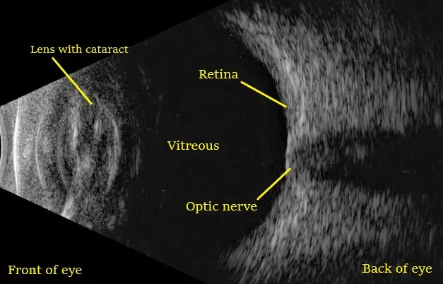
B-scan images are interpreted by the ophthalmologist to diagnose or assess various ocular conditions. Key structures that can be visualized include:
Vitreous Humor:
- The gel-like substance filling the eye, which may show floaters, hemorrhage, or other abnormalities.
Retina:
- The light-sensitive layer at the back of the eye, where conditions such as retinal detachment or tumors can be detected.
Choroid:
- The vascular layer of the eye, where choroidal detachments or masses can be identified.
Optic Nerve:
- The nerve transmitting visual information to the brain, where abnormalities like optic nerve head drusen or swelling can be observed.
Advantages of B-Scan Ultrasound
Non-Invasive:
- B-scan is a non-invasive procedure with no risk of radiation exposure, making it safe for repeated use.
Real-Time Imaging:
- Provides real-time visualization of the eye’s internal structures, allowing for dynamic assessment.
Versatility:
- Can be used in a variety of clinical situations, particularly when other imaging modalities are not feasible.
Limitations of B-Scan Ultrasound
Operator Dependency:
- The quality of the images and the diagnostic information obtained can vary depending on the skill and experience of the operator.
Limited Resolution:
- While B-scan provides valuable information, it may not offer the same level of detail as other imaging techniques, such as OCT (Optical Coherence Tomography) for certain structures.
Conclusion
B-scan ultrasound is a vital diagnostic tool in ophthalmology, particularly when direct visualization of the eye’s internal structures is not possible. It plays a critical role in diagnosing and managing conditions such as retinal detachment, vitreous hemorrhage, intraocular tumors, and foreign bodies. With its non-invasive nature and real-time imaging capabilities, B-scan continues to be an indispensable procedure in the assessment of ocular health.
USEFUL LINKS
SANKALP HOSPITALS PVT. LTD.
Vijay Smruti, Town Hall Circle, Jamnagar - 361 001 (Gujarat) INDIA
sankalphospitals@gmail.com
0288 2553435
© 2024 Sankalp Hospitals Pvt. Ltd.